What is lateral cephalometric analysis?
Lateral cephalometric analysis is a method of examining the skeleton of the face and jaws in numbers, using an X-ray of the head taken from the side (a lateral cephalogram).
It expresses the shape and position of the upper and lower jawbones, which lie behind the rows of teeth, as angles and lengths seen from the side.
Among our examination items, it corresponds to the "skeletal examination by X-ray", which also includes an orthopantomogram (panoramic X-ray).
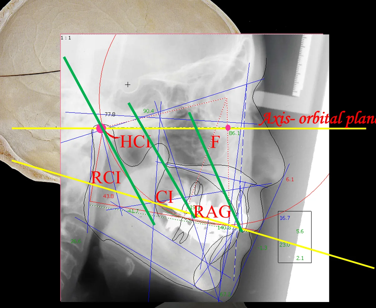
The photo shows our analysis screen.
Lines and symbols for analysing the skeleton are drawn over the X-ray of the head taken from the side.
How lateral cephalometric analysis works
The analysis generally proceeds in the following order.
- Taking the X-ray: an X-ray of the head is taken from the side.
- Marking points: defined points and outlines of the bones are specified on the image, on screen.
- Converting to numbers: values that describe the characteristics of the skeleton are calculated from the angles and lengths of the lines joining the points.
- Aligning the reference: at our clinic, we read the skeletal values on the same reference plane as the values for the jaw joints and the teeth.
Main values examined in the analysis
In our analysis, we look at values such as the following.
| Value or item examined | What it represents |
|---|---|
| Lower facial height (LFH) | The height of the lower part of the face |
| APDI | The front-to-back position of the lower jaw |
| Posterior discrepancy | The degree to which, in the back-tooth region, the bone supporting the teeth is insufficient for the size of the teeth |
| Vertical dimension of occlusion | The height between the upper and lower jaws when biting |
| Occlusal plane | The inclination of the surface where the upper and lower teeth meet |
The anteroposterior dysplasia indicator (APDI) was published in 1978 as an index that combines several angles on an X-ray of the head taken from the side to assess discrepancies in the front-to-back relationship between the upper and lower jaws.
At our clinic, we use it as a value for assessing the front-to-back position of the lower jaw.
Posterior discrepancy is a concept that Professor Sadao Sato of Kanagawa Dental University and colleagues have reported on since the 1980s.
Professor Sato and colleagues report that when the bony base in the back-tooth region is insufficient, the position of the back teeth and the inclination of the biting surfaces change, and that this also has a bearing on the position of the lower jaw.
The "HCI" on the screen is a Cadiax value
On the analysis screen in the photo, alongside the skeletal lines, there is also a line labelled "HCI".
HCI (horizontal condylar inclination) is the inclination of the path the jaw joint follows as it moves forward.
It is a value for jaw joint movement, not for the skeleton, and is recorded with Cadiax.
The method of viewing the skeletal analysis and the records of jaw joint movement superimposed on the same reference plane was introduced by Professor Rudolf Slavicek of the University of Vienna.
The manufacturer also states that its analysis program allows the analysis of X-rays of the head taken from the side to be viewed in connection with the records of jaw joint movement and with the articulator.
Using the same reference plane
The meaning of an angle or a length changes depending on what it was measured from.
A value needs a reference that tells you "where it is seen from".
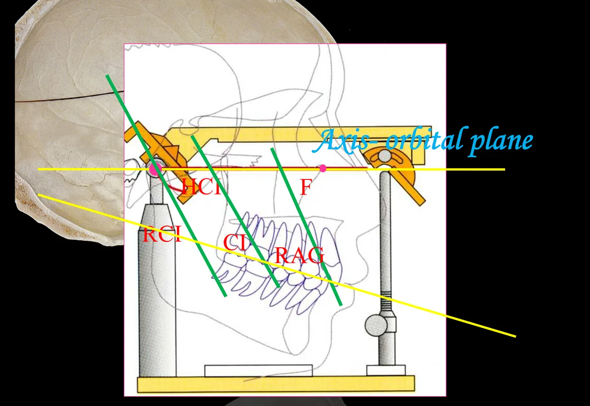
At our clinic, we measure the skeleton, jaw joint movement and tooth angles all from a single plane, called the axis-orbital plane.
The axis-orbital plane is the plane joining the axis of rotation of the left and right jaw joints (the hinge axis) and a point on the bone below the eye.
Values measured from the same reference can be represented as they are on the Girrbach Reference SL articulator.
This is why the results of the skeletal analysis can be carried through into the design of the bite on the casts.
Why we use lateral cephalometric analysis
Because the cause may lie in the skeleton itself
Some people with temporomandibular disorders (TMD) have well-aligned teeth.
When the cause lies in the skeleton itself, looking only inside the mouth, however closely, may not reveal any clue to a solution.
For this reason, at our clinic we look not only at the teeth but at the head as a whole, including the jaw joints and the skeleton.
Because we want to check the size difference between bone and teeth in numbers
The director believes that behind the problems with tooth alignment in people today lies a difference in size between bone and teeth.
As the director explains it, as humans moved to walking on two legs, the brain became larger, whereas the lower part of the face became smaller.
In this view, teeth that have not become much smaller are lined up in jawbones that have become smaller.
It can be compared to three large sumo wrestlers trying to sit together on a three-seater park bench.
With lateral cephalometric analysis, we check in numbers how much this difference (posterior discrepancy) is having an effect.
To measure on the same scale as the jaw joint and tooth values
When skeletal values are measured on the same reference plane as jaw joint movement and tooth angles, they can be brought together on the articulator.
We use the same reference so that the measured values can be given form as appliances and teeth.
To check how the skeleton has developed
This analysis also checks how the skeleton has grown.
For example, the analysis may reveal delayed development in the height of the lower part of the face, or insufficient development of the back parts of the upper and lower jaws.
In practice at our clinic
Its place among the examinations
Taking an X-ray of the head from the side is one of the detailed examinations carried out after the first consultation.
In the fee table, it is one of the items in the "orthodontic work-up (bite examination)" for TMD and orthodontic treatment.
The orthodontic work-up as a whole takes about 2–3 visits.
The combination of examinations, including whether to take the X-ray, is decided after looking at your symptoms and the condition of your mouth.
From X-ray to design
- As part of the detailed examinations, an X-ray of the head is taken from the side.
- Around the same time, as needed, we record jaw joint movement with Cadiax and examine each individual tooth using impressions and sectioned casts.
- We analyse the skeleton on the image and obtain values such as lower facial height and APDI.
- We align the skeletal values on the same reference plane as the jaw joint records and tooth angles, and decide the values to set on the articulator.
- We explain the results of the analysis using the numbers.
TMD and orthodontics
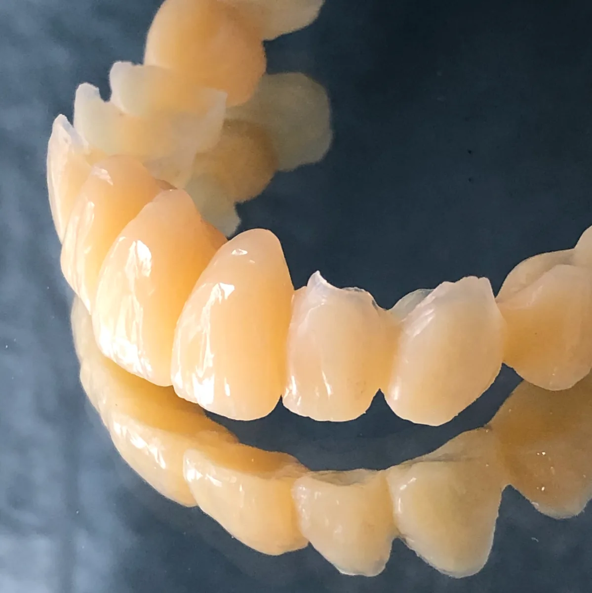
The characteristics of the skeleton are one of the things we consider when working out the target position of the lower jaw for treatment (the therapeutic jaw position).
That position is expressed in appliances such as metal overlays and provisional restorations, and checked in the actual mouth.
In orthodontic treatment, we combine the skeletal analysis with the records of jaw joint movement to plan how to move the teeth.
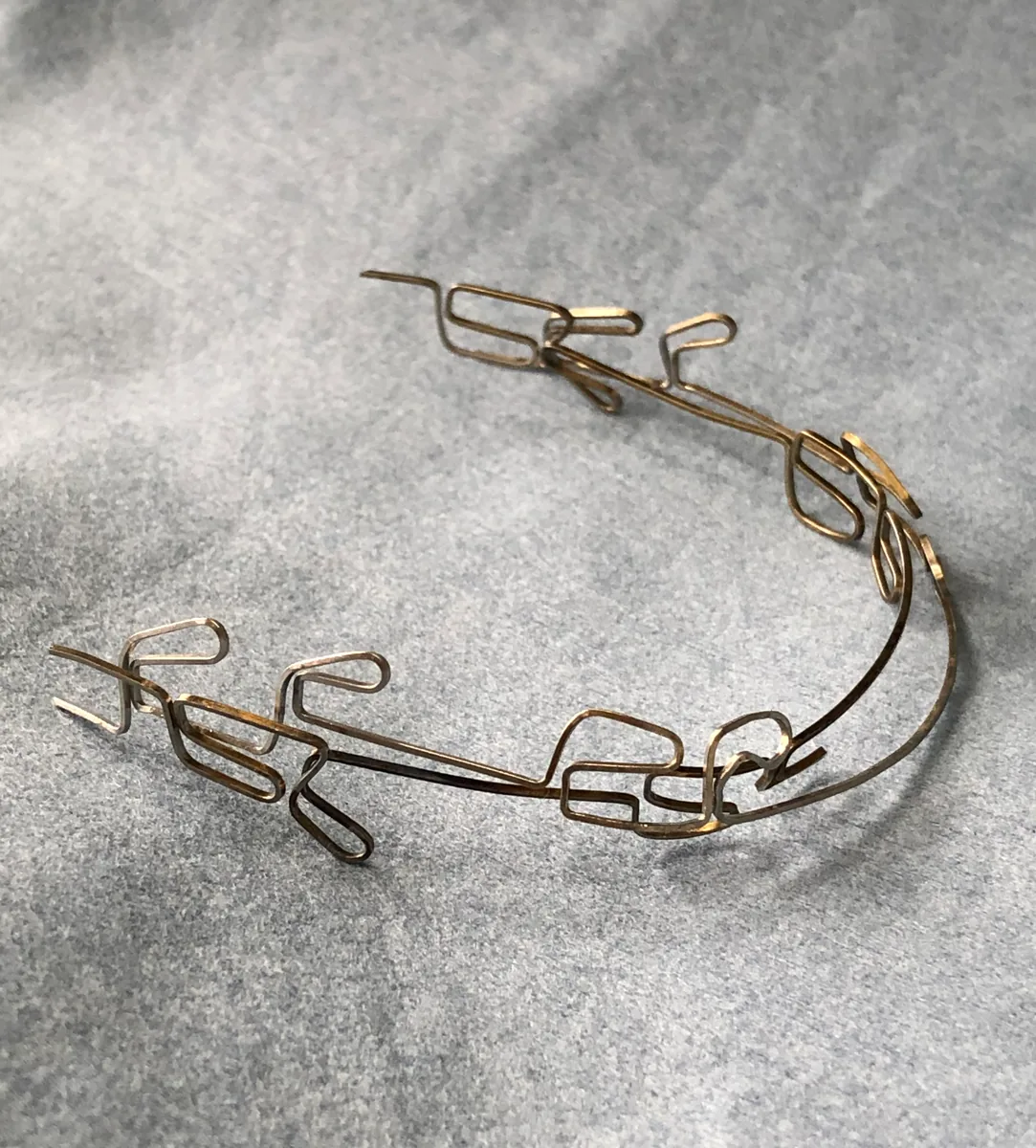
Dentures
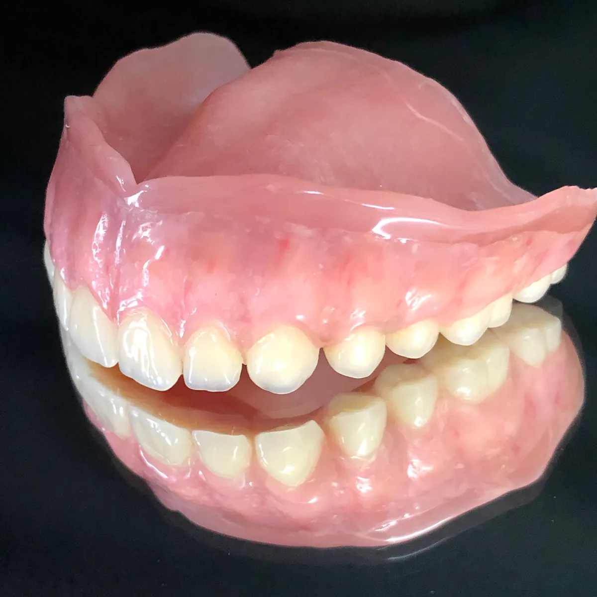
For complete dentures, the height between the upper and lower jaws when biting (vertical dimension of occlusion) and the inclination of the biting surfaces (occlusal plane) are decided based on the skeletal analysis.
The angle of the slopes of the artificial teeth (cusp inclination) is decided from the Cadiax records.
How the artificial teeth are arranged is explained on the artificial tooth arrangement page.
Crowns
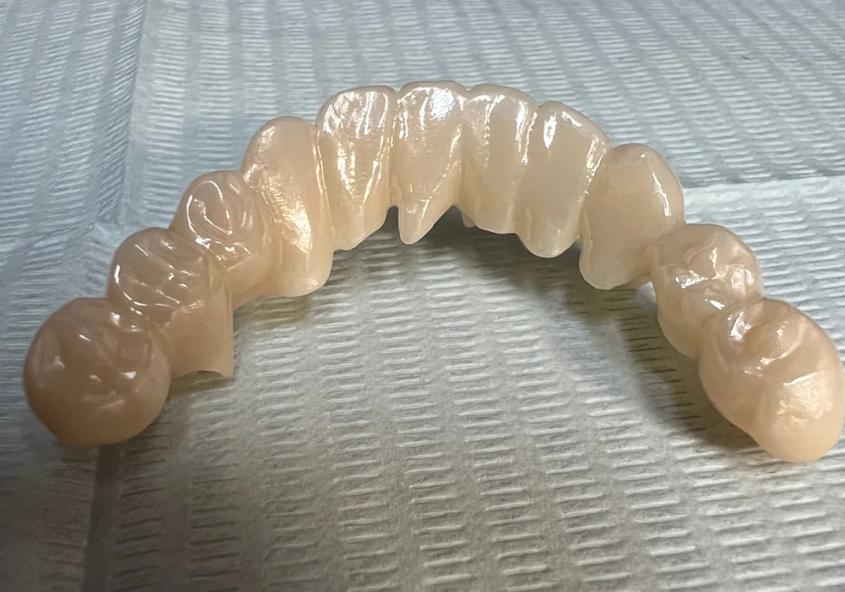
When many teeth are restored with crowns at the same time, too, the skeletal data, together with the jaw joint records and the analysis on the articulator, are used as a basis for deciding the shape.
What lateral cephalometric analysis can and cannot tell us
What lateral cephalometric analysis can tell us
- The positional relationship of the upper and lower jawbones, front to back and up and down
- The height of the lower part of the face (lower facial height)
- The effect of the size difference between bone and teeth (posterior discrepancy)
- The height between the upper and lower jaws when biting (vertical dimension of occlusion) and the inclination of the biting surfaces (occlusal plane)
- How the skeleton has developed
What it cannot tell us, and how we compensate
| What it cannot tell us | How we compensate |
|---|---|
| How the jaw joints actually move (including path angles such as HCI) | Cadiax |
| Traces of lower jaw movement while clenching | Intraoral gothic arch tracing (when making dentures) |
| The orientation and inclination of each tooth | Sectioned cast |
| The detailed inclination of the biting surfaces on casts of the dental arches | Occlusal plane measurement |
| Tooth contact during sleep | BruxChecker |
| Whether the position decided on the casts fits in the mouth | Checking while provisional restorations and appliances are in use (provisional restorations and provisional dentures) |
What an X-ray shows is the shape and positional relationships of the skeleton.
Why symptoms such as headaches and stiff shoulders are occurring cannot be determined from this analysis alone.
Depending on your symptoms, please also consider an assessment outside dentistry, such as in medicine.
Frequently asked questions
- Is this an examination of tooth alignment?
It examines not only tooth alignment but also the skeleton of the upper and lower jaws behind it.
It checks the relationship between the upper and lower jaws and the height of the lower part of the face as angles and lengths.
- Does this analysis also show jaw joint movement?
No. What this analysis shows is the shape and positional relationships of the skeleton.
Jaw joint movement (including path angles such as HCI) is recorded with Cadiax and viewed together with the skeletal analysis on the same reference plane.
- Why is a reference plane needed?
Because angles and lengths cannot be compared or combined unless the point they are measured from is the same.
At our clinic, we measure using the axis-orbital plane as the reference.
- Is the X-ray also taken when making dentures?
Yes. For complete dentures, it is used to decide the height between the upper and lower jaws when biting and the inclination of the biting surfaces.
- If I have the examination, does that mean I will have treatment?
No. Whether to carry out orthodontic or other treatment is decided after looking at the examination results, and in some cases it is not carried out.
After the examination, we tell you the possible options and the cost, duration and number of visits for each.
You can then consider whether to have treatment.
Related technologies and treatments
- Cadiax
- Girrbach Reference SL articulator
- Sectioned cast
- Austrian gnathology
- All the technologies we use
- Temporomandibular disorders & orthodontic treatment that avoids extractions where possible: examinations
- Dentures (precision dentures): examinations
- Thorough diagnosis before treatment (14 examination items)
Sources we referred to
For the general explanations (APDI, posterior discrepancy, HCI, the reference plane and how the analysis works), we referred to the following published sources. What we describe about our own methods is based on the director's posts and explanations.
- Kim YH, Vietas JJ. Anteroposterior dysplasia indicator: an adjunct to cephalometric differential diagnosis. Am J Orthod. 1978;73(6):619-633
- Sato S, Suzuki Y. Relationship between the development of skeletal mesio-occlusion and posterior tooth-to-denture base discrepancy. Nihon Kyosei Shika Gakkai Zasshi. 1988;47(4):796-810 (English abstract)
- Ahangari AH, et al. Evaluation of the Cadiax Compact II accuracy in recording preadjusted condylar inclinations on fully adjustable articulator. J Contemp Dent Pract. 2012;13(4):504-508
- GAMMA Dental, "Digital Diagnostic" (manufacturer)
- GAMMA Dental, "Face-bow transfer" (manufacturer)
- Kulmer S, et al. Incline and length of guiding elements in untreated naturally grown dentition. J Oral Rehabil. 1999;26(8):650-660
- Sukalo M. "Rudolf Slavicek und die Gnathologie in Österreich". Thesis, Medical University of Vienna, 2016 (in German; PDF)
Consultations and enquiries
Gibakai Medical Corporation
Yoshimi Dental Clinic
Square 2, Room 302 (third floor)
1-29-1 Tsukushino, Machida,
Tokyo 194-0001, Japan
TEL 042-795-1359 FAX 042-795-1398
Clinic days: Mon · Tue · Thu · Fri
10:00–13:00 / 14:30–18:00
A first visit begins with a first consultation (a private, non-insured service; 30 minutes to 1 hour) for examination and diagnosis. Questions about a diagnosis you received elsewhere are also covered within it.
In an emergency, such as being unable to chew or having severe symptoms, please call us on 042-795-1359 to discuss it.