What is a stereo microscope for laboratory work?
A stereo microscope for laboratory work is a microscope used in our in-house dental laboratory to work on and check provisional restorations, crowns, denture components, orthodontic wires and other items while viewing them magnified and in three dimensions.
It differs in both place and purpose from the dental operating microscope, which is used in the treatment room to view the area being treated under magnification.
This page describes the microscope in the dental laboratory.
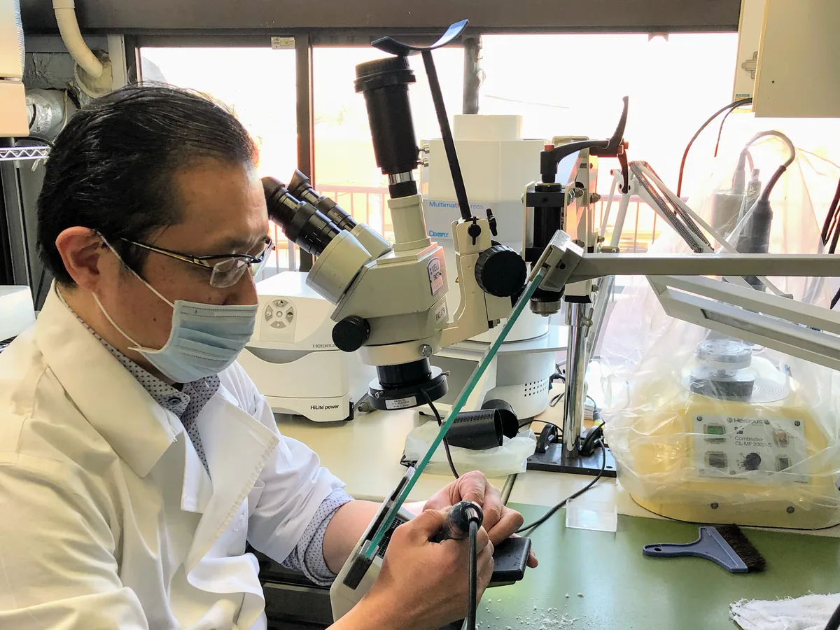
How a stereo microscope for laboratory work functions
Here we describe how a stereo microscope generally works, in four parts.
- Sending a separate image to each eye: the left and right eyes are sent images at slightly different angles.
- Seeing in three dimensions: the two images combine into one in the brain, so depth and surface relief are seen in three dimensions. This is the same mechanism by which people see objects in three dimensions with both eyes.
- Seeing in the same orientation as the object: because the image appears in the same orientation as the actual object, it is suited to work in which the hands move while looking.
- Allowing a wide gap between lens and object: because the gap between the lens and the object being viewed can be kept wide, it is easier to work with instruments while looking.
In general, the higher the magnification, the smaller the area that can be seen.
At the same time, the depth that appears in focus (depth of field) also becomes shallower.
In the microscope in our dental laboratory, the magnification of the eyepieces is deliberately kept low.
This is to increase the depth that appears in focus, making it easier to see the whole working area.
Why we use a stereo microscope for laboratory work
Because even small items have fine details of shape
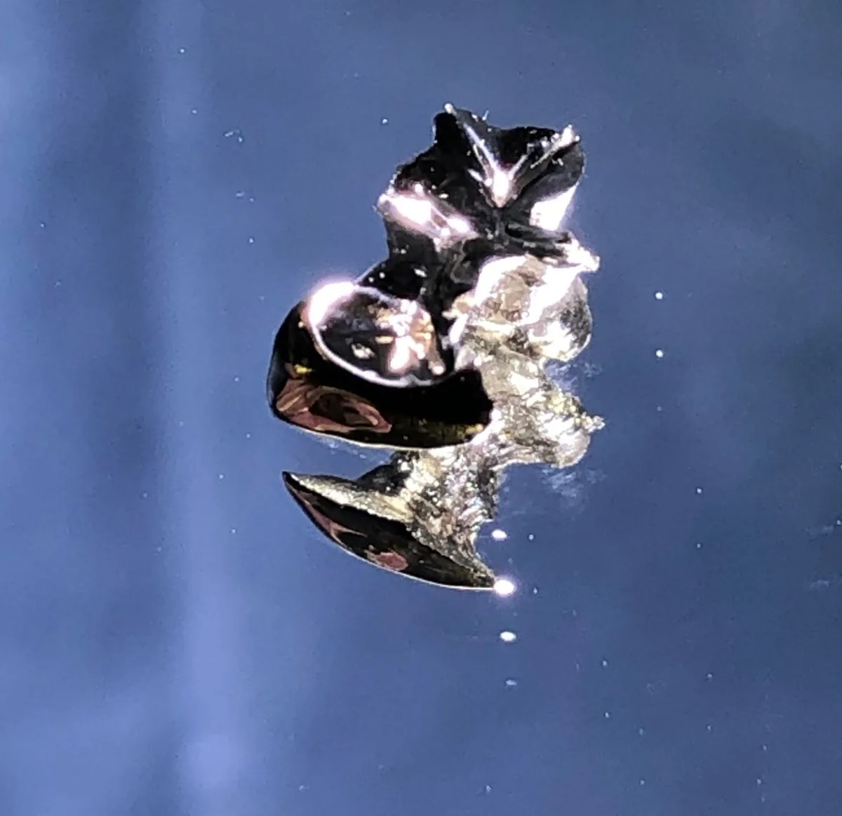
Crowns are small objects, but the director's view is that even within them, fine details of shape need to be created.
For this reason, we shape them while viewing them magnified and in three dimensions under the microscope in the dental laboratory.
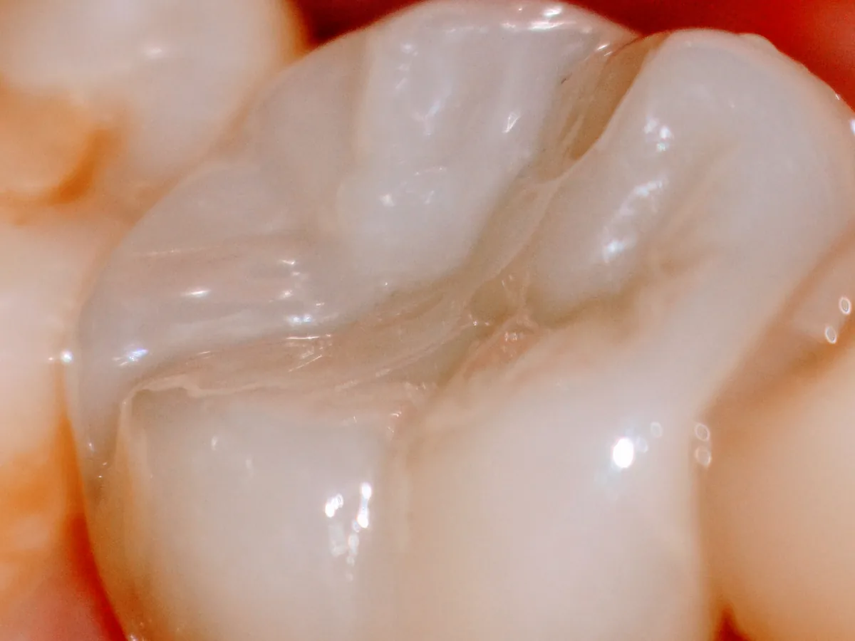
Because even a single provisional restoration is made while checking its shape
Even for a single small provisional restoration, we aim to shape it with its function and appearance in mind, in the same way as for the final crown.
We also use the microscope in the dental laboratory to make and check provisional restorations.
To give shape to the information from examinations
At our clinic, we place importance on expressing examination information, such as jaw movement, skeletal structure and tooth positions, in the shape of appliances and crowns.
The laboratory work that creates those shapes is carried out under the microscope.
In practice at our clinic
Which tasks we use it for
The microscope in the dental laboratory is used at the stages of creating the shape, joining metal and checking the finished item.
| Task | What we look at under the microscope | More details |
|---|---|---|
| Wax-up (making the shape in wax) | The shape of the teeth and of the bite being made on the cast | Wax-up |
| Making and checking provisional restorations | The shape and finish of small provisional restorations | Provisional restorations and provisional dentures |
| Checking finished crowns | The completed shape and finish | Zirconia crowns |
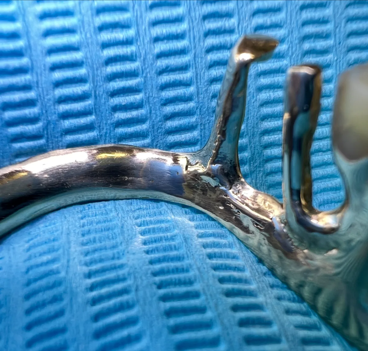
| Joining and repairs by laser welding | The part where metal is joined, and the shape around it | Laser welding |
| Checking orthodontic wires | The fine shape of the bent wire | MEAW |
| Fine adjustment of the devices in retainers | The fine details of the devices fitted in retainers | Retainer |
| Modifying plier tips | The shape of the laser-welded tips | Plier tip modification |
Orthodontic wires and the devices in retainers are adjusted in the dental laboratory next to the treatment room.
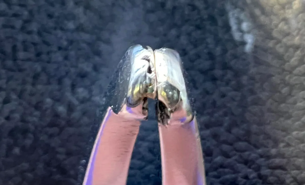
Combining it with other technologies
The microscope in the dental laboratory is not an instrument that decides the shape on its own.
The shape is based on records of jaw movement and skeletal structure, and on the articulator set up with them.
The microscope is used to refine that shape in detail and check the finish.
How it fits in the mouth is checked in the treatment room (see the "What it cannot do" table below).
What a stereo microscope for laboratory work can and cannot do
What it can do
- View provisional restorations, crowns, denture components and other items under magnification
- See depth and surface relief in three dimensions
- Move the hands and instruments while viewing an image in the same orientation as the object
- Check orthodontic wires and laser-welded parts
- Keep the eyepiece magnification low to see the whole working area
What it cannot do, and how we compensate
What can be seen with the microscope in the dental laboratory is the work outside the mouth.
How it fits in the mouth, how it relates to jaw movement and how it works in actual use cannot be checked in the dental laboratory alone.
Also, in general, the higher the magnification, the smaller the visible area and the shallower the depth in focus.
There are limits to making both the fine details and the whole working area easy to see at the same time at a single magnification.
| What it cannot do | How we compensate |
|---|---|
| Checking in the mouth how a crown fits the tooth | We check this with the dental operating microscope in the treatment room |
| Judging whether the work suits the bite and jaw movement | We work this out based on the Cadiax records and the articulator |
| Checking whether the position and shape are right in actual use | We check this while you use provisional restorations or provisional dentures |
| Seeing the whole working area while keeping the magnification high | We deliberately keep the eyepiece magnification low to increase the depth in focus |
Frequently asked questions
- How does it differ from the dental operating microscope in the treatment room?
The stereo microscope for laboratory work is an instrument used in our in-house dental laboratory to work on and check items being made while viewing them in three dimensions.
The dental operating microscope in the treatment room is used to view the area being treated inside the mouth under magnification.
- What kinds of items are viewed with it?
We look at provisional restorations, crowns, denture components, the devices in retainers, orthodontic wires, pliers and other items.
We check not only work in progress while the shape is being made, but also parts where metal has been joined and finished items.
- Why is the magnification deliberately kept low?
Because, in general, the higher the magnification, the smaller the visible area and the shallower the depth in focus.
At our clinic, we deliberately keep the eyepiece magnification low, making it easier to see the whole working area.
- Do you also make provisional restorations while looking through the microscope?
Yes.
Even for a single small provisional restoration, we aim to shape it with its function and appearance in mind, and we make and check it under the microscope.
- Does checking under the microscope show that it will fit in the mouth?
What the microscope in the dental laboratory can tell us is the shape and finish of the work.
How it fits in the mouth is checked with the dental operating microscope in the treatment room, and its relationship with the bite and jaw movement is checked using Cadiax, the articulator and provisional restorations.
Related technologies and treatments
- Dental operating microscope
- Wax-up
- Laser welding
- Provisional restorations and provisional dentures
- All the technologies we use
- Dentures (precision dentures)
- Aesthetic dentistry (crowns)
- Temporomandibular disorders & orthodontic treatment that avoids extractions where possible
Sources we referred to
For the general explanations (how things are seen in three dimensions, and the relationship between magnification, visible area and depth), we referred to the following published sources. What we describe about our own methods is based on the director's posts and the explanations on our current website.
- Nikon MicroscopyU, "Introduction to Stereomicroscopy" (manufacturer's explanatory article)
- Low JF, Mohd Dom TN, Baharin SA. Magnification in endodontics: A review of its application and acceptance among dental practitioners. Eur J Dent. 2018;12(4):610-616
Consultations and enquiries
Gibakai Medical Corporation
Yoshimi Dental Clinic
Square 2, Room 302 (third floor)
1-29-1 Tsukushino, Machida,
Tokyo 194-0001, Japan
TEL 042-795-1359 FAX 042-795-1398
Clinic days: Mon · Tue · Thu · Fri
10:00–13:00 / 14:30–18:00
A first visit begins with a first consultation (a private, non-insured service; 30 minutes to 1 hour) for examination and diagnosis. Questions about a diagnosis you received elsewhere are also covered within it.
In an emergency, such as being unable to chew or having severe symptoms, please call us on 042-795-1359 to discuss it.