What is a dental operating microscope?
A dental operating microscope is a microscope for dentistry, used to view the area being treated under magnification and illumination.
It is also called a dental microscope.
At our clinic, we use it for root canal treatment, checking crowns, checking provisional restorations and finished restorations, and periodontal treatment.
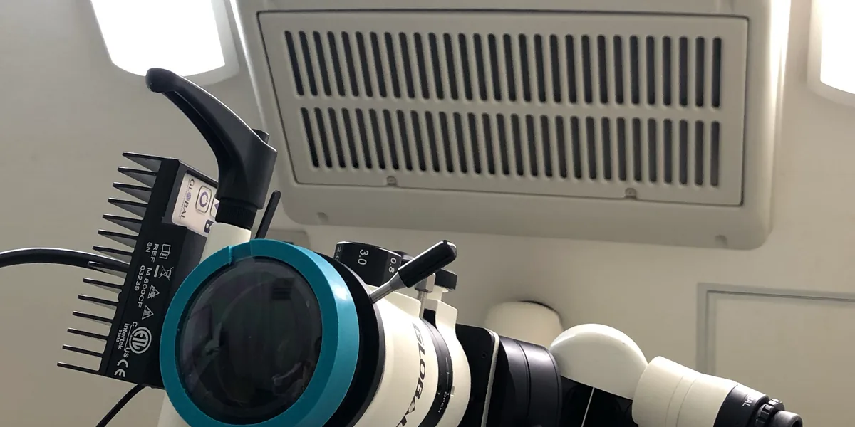
How a dental operating microscope works
Below, we describe how a typical dental operating microscope works, following the steps involved in viewing.
- Illuminating: light from the light source is directed through the objective lens onto the area being treated.
- Changing the magnification: the magnification is changed by turning a knob or using a zoom.
- Looking with both eyes: looking through the left and right eyepieces, the viewer sees the magnified area in three dimensions. The eyepieces can be adjusted to the viewer's eyesight.
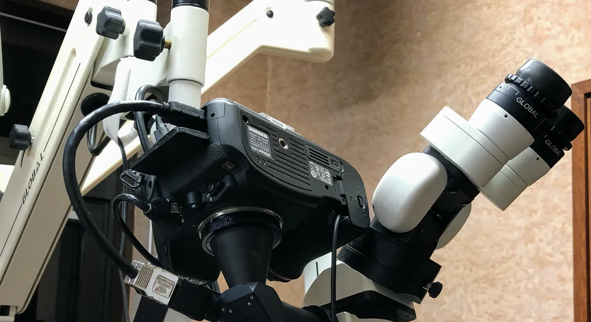
- Displaying and recording: with a camera connected, the image being viewed can be shown on a monitor or recorded as photos or video.
In general, increasing the magnification makes fine details look larger, but the area visible at one time becomes smaller and the depth that is in focus (depth of field) also becomes shallower.
For this reason, the magnification is generally switched to suit the purpose: low magnification to check the overall position, and high magnification to look at fine details.
At our clinic, we also display the image magnified by the microscope on the monitor used for treatment images, and check it there.
Why we use a dental operating microscope
Because we regard teeth as delicate sensors
The director regards the teeth not only as something for chewing food, but as sensors that pick up fine sensations.
The director believes that treating such teeth requires looking closely at the area being treated and building up fine procedures, and this is why we use the microscope.
Because there are fine structures inside the root
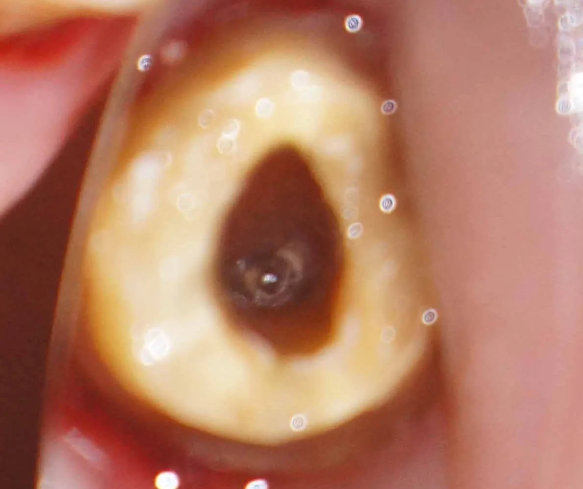
What root canal treatment has to deal with includes things that are hard to make out with the naked eye, such as cracks in the tooth and fine canals that branch off near the tip of the root (apical lateral canals).
We use the microscope to magnify and illuminate such areas and check them.
To check boundaries and deposits on the teeth
With crowns, we check under magnification the boundary with the tooth and how the crown fits.
In periodontal treatment, we remove plaque and tartar attached around the teeth while viewing them under magnification.
In practice at our clinic
Which treatments, and what we look at
We use it according to the type of treatment and what we want to check.
| Treatment | When we use it | What we look at |
|---|---|---|
| Root canal treatment | When treating inside the root canal and checking the condition of the root tip | The shape of the root canal, the opening at the root tip, cracks in the tooth and so on |
| Crowns | When checking how a crown fits | The boundary between the crown and the tooth, and whether there is a gap there |
| Provisional restorations and finished restorations | When checking provisional restorations and finished restorations | Shape and finish |
| Periodontal treatment | When removing deposits from around the teeth | Plaque and tartar |
In root canal treatment
"Microscopic endodontics" is the name given to root canal treatment carried out while directly viewing, through the microscope, the opening at the end of the root canal (the apical foramen).
The steps of root canal treatment are explained in "How we carry out root canal treatment" on the root canal treatment page.
For crowns, provisional restorations and finished restorations
For crowns, we check the fit under magnification to see whether there is any gap between the crown and the tooth.
Provisional restorations and finished restorations are also checked with the microscope.
What the microscope can tell us is the shape and boundaries of the magnified area.
Whether a crown suits the bite and jaw movement is worked out based on records of jaw movement and the articulator (see the "What it cannot tell us" table below).
How it differs from the microscope in the dental laboratory
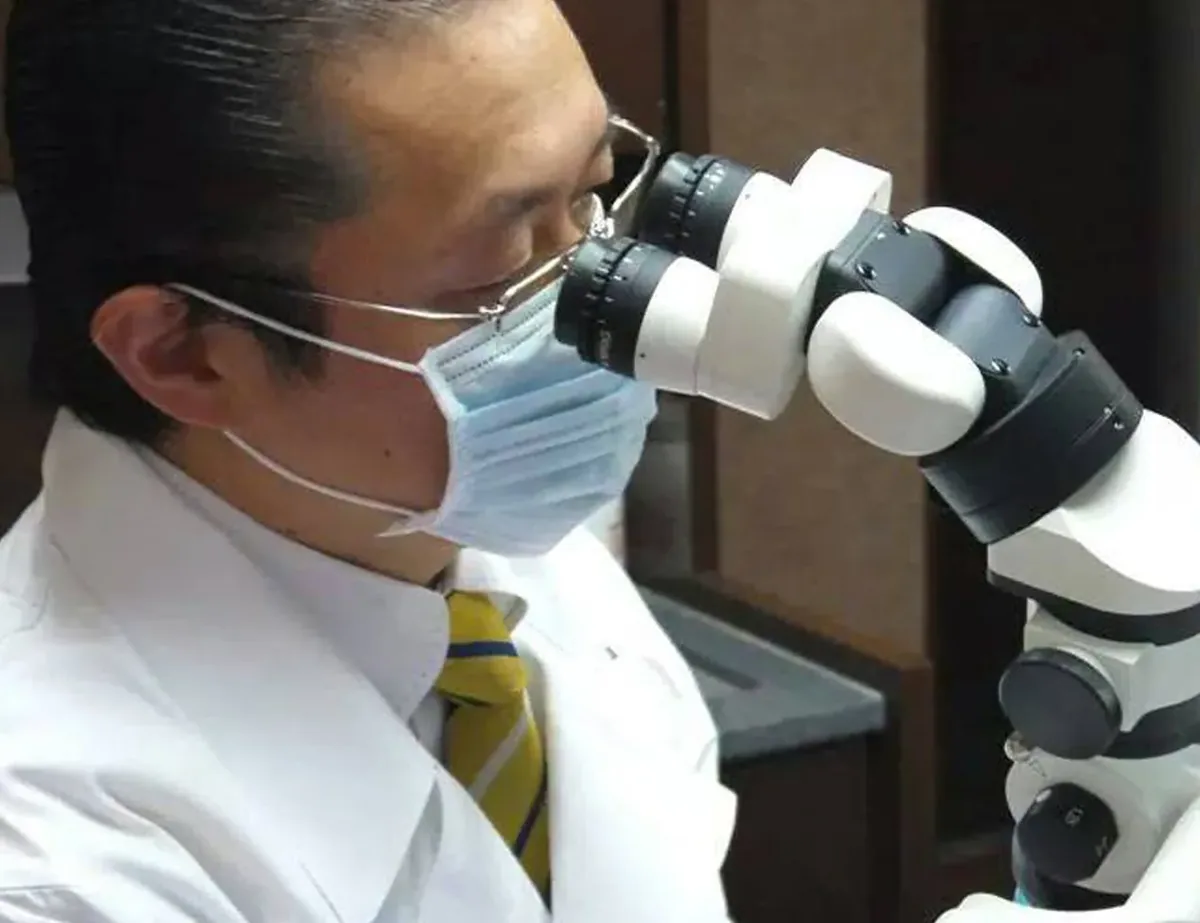
The microscope on this page is an instrument for viewing the area being treated inside the mouth.
The microscope used outside the mouth to work on laboratory-made items while viewing them magnified is described on the stereo microscope for laboratory work page.
What a dental operating microscope can and cannot tell us
What a dental operating microscope can tell us
- A magnified, three-dimensional image of the illuminated area being treated
- Fine shapes within the visible range, such as root canal entrances, the shape of the root canals and cracks in the teeth
- The boundary between a crown and the tooth, and how the crown fits
- Plaque and tartar attached around the teeth
What it cannot tell us, and how we compensate
A microscope is an instrument for seeing.
In general, the higher the magnification, the smaller the area visible at one time.
It has also been reported that, with lower back teeth and in similar cases, getting the right viewing angle and posture can be difficult.
Being able to see under magnification does not guarantee that the tooth can be kept, or how things will go after treatment.
Results vary from person to person.
Depending on the condition of the root canals or cracks, retreatment or extraction may be needed.
Where looking alone cannot tell us something, we compensate as follows.
| What it cannot tell us | How we compensate |
|---|---|
| Whether a crown suits the bite and jaw movement | We record jaw movement with Cadiax and decide the shape based on the articulator |
| The fine shape of laboratory-made items outside the mouth | We check it with the stereo microscope for laboratory work |
| Whether the shape decided on fits in actual use | We check this while you use a provisional restoration |
Frequently asked questions
- Is a dental operating microscope an electron microscope?
No.
It is a microscope that uses lenses and light to view the area being treated under magnification.
With a camera attached, the image being viewed can also be shown on a monitor.
- Is it used only for root canal treatment?
No.
At our clinic, besides root canal treatment, we also use it to check how crowns fit, to check provisional restorations and finished restorations, and in periodontal treatment.
- Can the image seen through the microscope also be shown on a monitor?
Yes.
At our clinic, we also display the magnified image on the monitor used for treatment images, and check it there.
- Is there anything the microscope alone cannot tell us?
Yes, there is.
What the microscope can tell us is the shape and boundaries of the magnified area.
Whether a crown suits the bite and jaw movement is worked out based on the Cadiax records and the articulator.
- Does using a microscope mean the tooth can be kept?
A microscope is an instrument for checking the shape of the root canals, the root tip, cracks and so on under magnification.
What we learn from it becomes a basis for planning treatment that aims to keep the tooth.
However, the outlook differs depending on the condition of the root canals and cracks, and retreatment or extraction may become necessary.
Related technologies and treatments
- Stereo microscope for laboratory work
- Cadiax
- Girrbach Reference SL articulator
- Provisional restorations and provisional dentures
- All the technologies we use
- Root canal treatment
- Aesthetic dentistry (crowns)
- Periodontal disease
Sources we referred to
For the general explanations (how illumination, magnification, viewing with both eyes and recording work) and how the view changes with magnification, we referred to the following published sources. What we describe about our own methods is based on the director's posts and the explanations on our current website.
- Cordero I. Understanding and caring for an operating microscope. Community Eye Health. 2014;27(85):17
- Low JF, Mohd Dom TN, Baharin SA. Magnification in endodontics: A review of its application and acceptance among dental practitioners. Eur J Dent. 2018;12(4):610-616
- Chauhan S, et al. Magnification: The game changer in dentistry. World J Methodol. 2025;15(2):100937
- American Association of Endodontists, "The Dental Operating Microscope in Endodontics". Endodontics: Colleagues for Excellence, Winter 2016 (PDF)
Consultations and enquiries
Gibakai Medical Corporation
Yoshimi Dental Clinic
Square 2, Room 302 (third floor)
1-29-1 Tsukushino, Machida,
Tokyo 194-0001, Japan
TEL 042-795-1359 FAX 042-795-1398
Clinic days: Mon · Tue · Thu · Fri
10:00–13:00 / 14:30–18:00
A first visit begins with a first consultation (a private, non-insured service; 30 minutes to 1 hour) for examination and diagnosis. Questions about a diagnosis you received elsewhere are also covered within it.
In an emergency, such as being unable to chew or having severe symptoms, please call us on 042-795-1359 to discuss it.