The relationship between occlusion and the autonomic nervous system
Occlusion (the bite) is one of the factors that influence the physiological functions of the mandibular-cranial system.
Our clinic researches the relationship between occlusion and the whole body (this research does not indicate treatment outcomes for individual patients). Autonomic nervous function plays a key role in maintaining human homeostasis, and changes in occlusal condition brought about by dental treatment can be expected to act on autonomic function and affect the maintenance of emotion and cognitive function. At present, however, there are few reports on differences in autonomic function according to differences in occlusal pattern.
Yoshimi Dental Clinic focused on this point. We fitted various occlusal-surface metal overlays onto the dental arch and measured changes in autonomic nervous function immediately after several minutes of grinding movement, using the pupillary reflex. The pupillary reflex is measured non-invasively with the Iriscorder Dual C10641 pupillometer to obtain data on changes in autonomic function. Electrocardiogram and electromyogram data are collected as well, with the aim of comprehensively investigating changes in the occlusal surface morphology of the dental arch and changes in autonomic nervous function.
Background to our focus on
the link with autonomic function


There are many reports demonstrating the existence of a trigeminal-autonomic reflex in the craniomandibular system. It is an important reflex involved in regulating the metabolism of organs throughout the body. Its existence therefore cannot be ignored when treating the stomatognathic and craniomandibular system. Our clinic also focuses its research on the relationship between the mouth and the whole body.
To break through this situation, the influence of the oral cavity on the autonomic centers needs to be clarified more sharply. In other words, we believe it is important to define the therapeutic goal of dentistry clearly. The pupillary reflex has the advantage of allowing autonomic function to be evaluated quickly and non-invasively, and is a physiological test well suited to future application as a clinical examination.
Our hypothesis is that if changes in peripheral occlusal condition appear as changes in autonomic activity, then pupillary reflex function, which is under autonomic control, will also change. Using this pathway from trigeminal stimulation to changes in pupil diameter, we hope to clarify the influence of occlusal changes on autonomic nervous function.
Contribution to future
health maintenance and promotion
Clarifying the influence of different occlusal patterns on autonomic nervous function will make it possible to set clear target values in clinical fields dealing with the oral region, such as prosthodontics, orthodontics, conservative dentistry and oral surgery.
Application to managing the growth and development of children is also a subject of our research.
130th Anniversary Scientific Meeting of
the Japan Prosthodontic Society
I took part in the symposium "Update on Occlusion Theory" at the 130th Anniversary Scientific Meeting of the Japan Prosthodontic Society. At the symposium I presented on "The Importance of Objective Examination and Diagnosis in Occlusal Reconstruction and the Potential of Occlusal Treatment," suggesting that occlusal treatment may improve responses in the limbic system, a higher center of the autonomic nervous system.
The Importance of Objective Examination and Diagnosis in Occlusal Reconstruction and the Potential of Occlusal Treatment

In medicine, treatment plans are decided on the basis of objective data such as body temperature, X-rays and blood tests. Our clinic likewise decides treatment plans on the basis of pre-operative examination data. Professor Emeritus Rudolf Slavicek of the Department of Prosthodontics, University of Vienna, developed devices for measuring objective data in occlusal reconstruction and established the discipline of occlusal reconstruction (Austrian gnathology). From 2000, under Professor Emeritus Sadao Sato of the Department of Craniofacial Growth and Development Dentistry, Kanagawa Dental University, I was responsible for clinical treatment based on this theory (orthodontics, prosthodontics and TMD treatment) and clinical research (development and analysis of equipment for recording sleep bruxism data). I believe that deciding the treatment plan and the specific means of treatment on the basis of objective data can reduce differences between practitioners. As the sleep experiments progressed, it became clear that there is a strong relationship between sleep bruxism and heart rate variability. Autonomic analysis data obtained using the precise analytical techniques of the engineering laboratory to which I currently belong showed changes in autonomic activity at each step of occlusal treatment. Based on this experience and these results, I will discuss the potential of occlusal treatment.
Topics
1. Objective examination and diagnosis.
2. Scientific occlusal reconstruction methods.
3. Occlusion and autonomic reflexes.
Reference literature
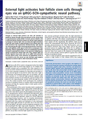
External light activates hair follicle stem cells through eyes via an ipRGC-SCN-sympathetic neural pathway.
This paper shows that light stimulation to the eyes activates hair follicle stem cells involved in hair regeneration via the ipRGC-SCN autonomic circuit. The mechanism is that the light signal is transmitted via melanopsin to the SCN, after which the sympathetic nervous system is immediately activated and releases norepinephrine, which in turn activates the hair follicle stem cells.
Since sympathetic activation is not limited to the skin, light stimulation to the eyes is expected to promote activation of other tissues as well.
To help maintain our patients' health, we will continue to comprehensively investigate occlusion and changes in autonomic nervous function.