What is pupillary light reflex measurement?
Pupillary light reflex measurement is a method that records the change in pupil size when light is shone into the eye and expresses it as numbers.
At our clinic, we use the Iriscorder Dual (C10641), a pupillometer made by Hamamatsu Photonics.
The pupil is the part in the centre of the coloured part of the eye that lets light through.
When light enters the eye, the pupil becomes smaller and then returns to its original size.
This response is called the pupillary light reflex, or light reflex.
Its generic name as a medical device is "pupillometer".
Among our examination items, this corresponds to "autonomic reflex testing by pupillary light reflex".
We use pupillary light reflex measurement in research investigating the relationship between occlusion and autonomic nervous function.
This page introduces a measurement technique; it does not describe the effects of treatment.
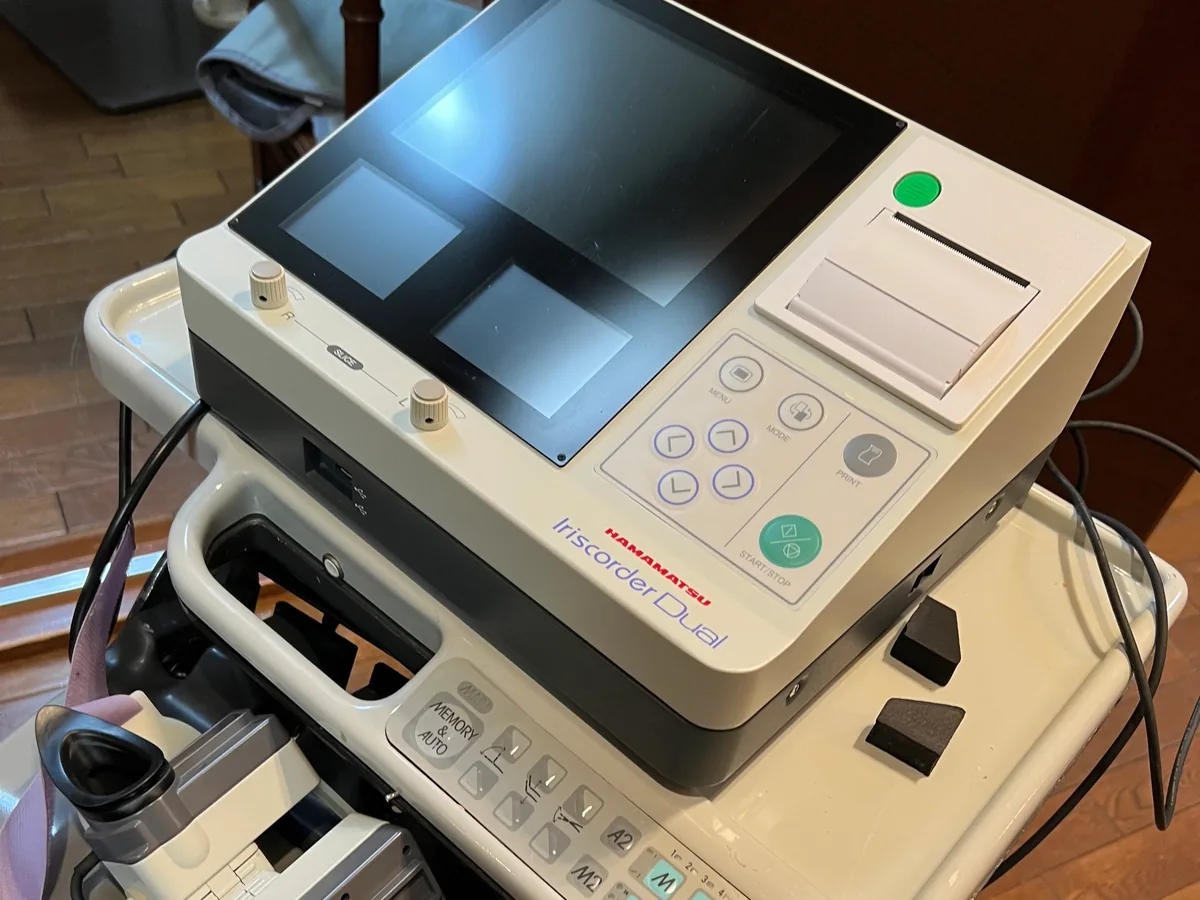
Background to its development
The Iriscorder is a device that Hamamatsu Photonics began developing in 1969 at the request of Satoshi Ishikawa, then a lecturer in the Department of Ophthalmology, Faculty of Medicine, University of Tokyo.
The aim was to measure accurately the response of the pupil (the change in pupil area), which is closely linked to the workings of the autonomic nervous system.
How pupillary light reflex measurement works
Imaging the pupil with infrared light
Based on the description in the marketing authorization holder's electronic package insert, we explain how it works in four steps.
- The eye is illuminated with infrared light and imaged with an infrared camera. Because the human eye does not perceive infrared light and the pupil does not respond to it, the illuminating light does not change the size of the pupil.
- Because the illuminating light is not reflected at the pupil, the pupil appears darker than the rest of the eye. This dark area is extracted, and the horizontal length of the pupil (horizontal diameter) is measured.
- A specified visible light is shone briefly into the eye, and the way the pupil responds is recorded (measurement of the light response). The light is shone for about 1 second.
- From the recording, values such as the size of the pupil and the speed and timing of the response are calculated.
According to the electronic package insert, the intensity of the light used is classified in the "Exempt Group" of the international standard on the effects of light on the eye (IEC 62471).
How the pupil becomes smaller and larger
The pupil becoming smaller is called "miosis", and the pupil becoming larger is called "mydriasis".
The parasympathetic nerves are involved in miosis, and the sympathetic nerves in mydriasis.
The action that makes the pupil smaller is transmitted through the nerve that moves the eye (the oculomotor nerve).
The action that makes it larger is transmitted through a sympathetic pathway that begins in the hypothalamus of the brain.
The parasympathetic and sympathetic nerves are both part of the autonomic nervous system.
Recording the pupillary light reflex makes it possible to see one of the responses involving the autonomic nervous system as numbers.
Iriscorder Dual
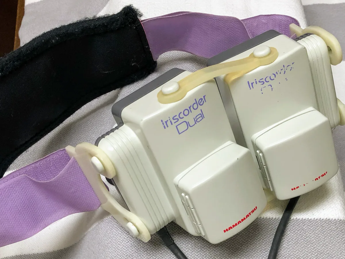
Recording the left and right eyes at the same time
The Iriscorder Dual is a measuring instrument that can record the pupils of the left and right eyes at the same time.
Measurements are taken with the goggle-shaped measuring unit placed over both eyes.
In our research, we used red light (wavelength 635 nm) and blue light (wavelength 470 nm).
Red light is the light used as standard to elicit the pupillary light reflex.
The instrument does not touch the eyes directly (non-contact), and the measurement is made without injuring the body (non-invasive) and takes a short time.
How to read the recording screen
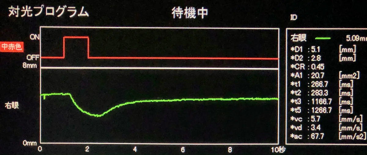
The red line at the top of the recording screen shows the period during which the light is shone.
The green line below is the diameter of the pupil.
When the light is shone, the line goes down: the pupil becomes smaller and then gradually returns to its original size.
From the recording, we obtain the following values.
| Symbol | Meaning |
|---|---|
| D1 | Diameter of the pupil before the light is shone |
| D2 | Diameter of the pupil at its smallest after the light is shone |
| CR | Proportion by which the pupil became smaller (constriction ratio) |
| t1 | Time from when the light is shone until the pupil begins to become smaller |
| t2 | Time from when the pupil begins to become smaller until it reaches half (50%) of the total constriction |
| t3 | Time until the pupil is at its smallest |
| t5 | Time from when the pupil is at its smallest until its diameter has recovered by 63% |
| vc | Maximum velocity while the pupil becomes smaller |
| vd | Maximum velocity while the pupil becomes larger |
| ac | Maximum acceleration while the pupil becomes smaller |
These values are shown on the recording screen.
In our research, these values were calculated from the recorded pupil diameter data using a program written for the research (MATLAB).
Why we measure the pupillary light reflex
To study the relationship between occlusion and the autonomic nervous system in a measurable form
A nerve called the trigeminal nerve is involved in the sensation and movement of the upper and lower jaws and the teeth.
Many reports have been published on reflexes between the trigeminal nerve and the autonomic nervous system in the region around the jaws.
On the other hand, we consider that there have been few reports on how autonomic nervous function differs with differences in occlusion.
The director focused on this point and, since 2002, has been investigating it in a measurable form.
Why we look at the pupillary light reflex
The trigeminal nerve, which carries sensation from the teeth and jaws, is not directly part of the neural circuit of the pupillary light reflex.
The paper explains that sensation from the mouth and face is linked to the state of the autonomic nervous system through the workings of the hypothalamus, and may appear indirectly in the movement of the pupil.
In our research, we chose the pupillary light reflex, which can be measured in a short time, in order to record the response immediately after tooth grinding.
It is said that there are still few studies that have examined the pupillary light reflex in relation to occlusion or tooth grinding.
Because we want to make treatment goals clear with data
The director wants to make clear, from measured information, what dental treatment should aim for.
He also believes that deciding the treatment plan on the basis of objective data can reduce differences between practitioners.
In practice at our clinic
How it was measured in the research
In the study reported in the 2026 paper, the pupillary light reflex was measured in the following order.
- The participant sits on a chair in a dark, quiet room for 20 minutes to let the eyes adjust to the dark (dark adaptation). At the end of this period, a baseline pupillary light reflex is recorded.
- The head is supported by a chin rest and a forehead rest, and the goggle-shaped measuring unit is placed over both eyes.
- The occlusal condition is changed temporarily with a removable metal appliance worn on the teeth (a metal overlay), and the participant makes tooth-grinding movements, moving the lower jaw from side to side, for 30 seconds.
- Immediately afterwards, red light and blue light are each shone for 1 second, and the pupil diameter is recorded 60 times per second from 1 second before to 10 seconds after the light.
- After a 2-minute rest, the appliance is changed and the same measurement is made under the other occlusal condition.
The participants were 17 healthy adults.
The study was approved by the ethics review committees of Gibakai Medical Corporation and Meiji University, and was carried out with written consent from the participants.
The recordings are analysed in the laboratory of Professor Yumie Ono, School of Science and Technology, Meiji University, to which the director belongs.
Research using the pupillary light reflex was also presented at the meetings of the Japan Society of Autonomic Neuroscience in 2019 and 2020.
The 2020 presentation dealt with the pupillary light reflex when red light was shone.
The presentations and papers are listed on the Research, publications and conferences page.
Use in clinical practice
In clinical practice, we may measure the pupillary light reflex for patients for whom we judge it necessary.
It is not an examination we carry out for every patient; we choose the examinations needed according to symptoms and condition.
We also show patients their examination results.
Examination duration, number of visits and fees → Fees
Combining it with other examinations
In detailed examinations for temporomandibular disorders (TMD), we combine, as needed, recording of jaw movement (Cadiax), skeletal analysis, electromyography, autonomic reflex testing and other examinations.
In our research, we also record the electrocardiogram (heart rate variability) and the activity of the chewing muscles (electromyography) on the same occasion as the pupillary light reflex.
What pupillary light reflex measurement can and cannot tell us
What it can tell us
- The change in pupil size when light is shone (the diameter, and the proportion by which the pupil became smaller)
- The time from when the light is shone until the response begins
- The speed and timing of the pupil becoming smaller and returning to its original size
- The responses of the left and right eyes (both eyes recorded at the same time)
- Differences between recordings made by the same method under different conditions
What it cannot tell us, and how we compensate
| What it cannot tell us | How we compensate |
|---|---|
| Changes in the autonomic nervous system over time | Heart rate variability (in our research, the electrocardiogram was recorded during tooth grinding) |
| How much the chewing muscles worked | Electromyography |
| Jaw joint movement | Cadiax |
| The cause of symptoms such as headaches, stiff shoulders and sleep problems | We do not determine it from pupillary light reflex values or the bite alone. Where needed, we recommend assessment by a physician |
What pupillary light reflex values show is the pupil's response at that moment, when light is shone under defined conditions.
What the paper examined was the response of healthy adult participants immediately after they ground their teeth with the occlusal condition temporarily changed.
The paper states that further research is needed on the relationship with occlusal conditions that have persisted for a long time.
The associations examined in research are separate from the reasons each person's symptoms occur and from the effects of their treatment.
Pupillary light reflex measurement is not for improving symptoms; it is for recording responses.
Frequently asked questions
- Why do you measure the pupillary light reflex at a dental clinic?
We research the relationship between occlusion and autonomic nervous function.
Because the parasympathetic and sympathetic nerves are involved in the movement of the pupil, we use it as one method of recording responses that involve the autonomic nervous system.
- Does the instrument touch the eyes or injure the body?
The instrument does not touch the eyes directly.
The goggle-shaped measuring unit is placed over both eyes, and the pupils are imaged with an infrared camera.
It is a measurement made without injuring the body (non-invasive).
- What kind of light is shone into the eyes?
A specified visible light is shone for about 1 second.
In the study reported in the 2026 paper, red light and blue light were used.
The photo of the recording screen on this page was taken when red light was shone.
- What do the values on the recording screen represent?
They include the diameter of the pupil before the light and at its smallest, the time until the pupil begins to become smaller, and the speed at which it constricts and recovers.
All of them are values that describe the pupil's response at the time of measurement.
- Can pupillary light reflex values show the cause of symptoms?
The cause of symptoms cannot be determined from pupillary light reflex values alone.
Where needed, we also recommend assessment by a physician.
Related technologies and treatments
- Heart rate variability (HRV)
- Electromyography (EMG)
- Cadiax
- Metal overlay
- All the technologies we use
- Temporomandibular disorders & orthodontic treatment that avoids extractions where possible: headaches, stiff shoulders, tinnitus and difficulty sleeping
- Occlusion and autonomic nervous function
- Research, publications and conferences
Sources we referred to
For the general explanations (how it works and the background to its development), we referred to the following published sources. What we describe about our own methods is based on the director's posts, papers and explanations.
- Iriscorder Dual C10641 electronic package insert (public document of the Pharmaceuticals and Medical Devices Agency, Japan; in Japanese)
- Hamamatsu Photonics, "[History of Hamamatsu Photonics]" (in Japanese)
- Yoshimi H, Suzuki T, Ono Y. Autonomic Responses to Tooth Grinding under Different Dental Occlusal Conditions. Advanced Biomedical Engineering. 2026;15:36-45
Consultations and enquiries
Gibakai Medical Corporation
Yoshimi Dental Clinic
Square 2, Room 302 (third floor)
1-29-1 Tsukushino, Machida,
Tokyo 194-0001, Japan
TEL 042-795-1359 FAX 042-795-1398
Clinic days: Mon · Tue · Thu · Fri
10:00–13:00 / 14:30–18:00
A first visit begins with a first consultation (a private, non-insured service; 30 minutes to 1 hour) for examination and diagnosis. Questions about a diagnosis you received elsewhere are also covered within it.
In an emergency, such as being unable to chew or having severe symptoms, please call us on 042-795-1359 to discuss it.