What is electromyography?
Electromyography (EMG) records, as waveforms, the electrical changes that occur when muscles work.
In our research, we record the activity of the cheek muscle that works in chewing (the masseter muscle) from the surface of the skin.
Among our examination items, this corresponds to "muscle activity testing by electromyography".
Where needed, electromyography is also carried out in detailed examinations for temporomandibular disorders (TMD).
This page describes how electromyography recording works and how it is used.
It is not a page that describes the effects of treatment.
How electromyography works
Recording electrical changes in the masseter muscle from the surface of the skin
Based on the study reported in the 2026 paper, we describe how the recording is made.
- The skin over the left and right masseter muscles is wiped with an alcohol swab. This is to prevent unwanted signals (noise) from entering the recording.
- Two electrodes are attached as a pair over the masseter muscle. The method of recording muscle activity from the surface of the skin is called surface electromyography (surface EMG).
- First, three recordings are taken of clenching with maximum force for 7 seconds.
- Using the largest value in those recordings as the reference, the activity of the masseter muscle at other times is expressed as a percentage (%MVC, percentage of maximal voluntary contraction).
| When it is recorded | How it is expressed |
|---|---|
| Clenching with maximum force for 7 seconds (3 times) | Used as the reference for comparison |
| While relaxed and at rest | Percentage of the reference (%MVC) |
| While making tooth-grinding movements under changed occlusal conditions | Percentage of the reference (%MVC) |
The paper states that using maximum clenching as the reference is a method commonly used in electromyography recording.
This is because clenching tends to produce similarly large values each time, whereas tooth grinding, which involves large movements, is not suited to serving as the reference.
Recording at the same time as the electrocardiogram
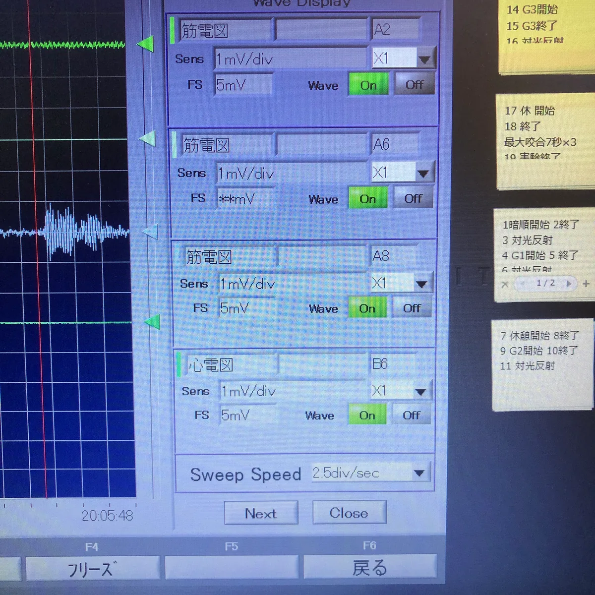
Settings screen for electromyography and electrocardiography
In our research, electromyography is recorded at the same time as the electrocardiogram, using the same multichannel recording device (a telemetry system).
The aim is to collect electrocardiogram and electromyogram data together and examine them comprehensively.
The screen in the photo shows three electromyography settings and one electrocardiography setting.
Why we use electromyography
To check the work of the chewing muscles with recordings
The director considers electromyography and electrocardiography necessary as part of the examinations for understanding the current state of TMD.
We check, as waveforms and numbers, when and how much the chewing muscles worked.
To see how muscle activity differs with occlusal conditions
In the 2026 study, electromyography was recorded to see whether masseter muscle activity differed when tooth-grinding movements were made under changed occlusal conditions.
To study tooth grinding during sleep
At Kanagawa Dental University, the director was responsible for developing equipment to collect data on tooth grinding during sleep (sleep bruxism) and for analysing those data.
Electromyograms of the masseter muscle have also been recorded in the research on tooth grinding during sleep.
In practice at our clinic
Recording in research
In the study reported in the 2026 paper, after maximum clenching had been recorded, 17 healthy adult participants made tooth-grinding movements for 30 seconds under each of two different occlusal conditions.
During this time, electromyograms of the left and right masseter muscles were recorded together with the electrocardiogram.
Recording tooth grinding during sleep
In a 2009 paper with the director as first author, a recording system combining an accelerometer that records movement in two directions, electroencephalography and electromyography was built to study tooth grinding during sleep.
Recordings were made of 19 healthy participants in a sleep laboratory.
The paper reports that this recording made it possible to distinguish tooth-grinding movements by type: rubbing the teeth together, clenching, and tapping the teeth together.
In the study reported in the 2018 paper, too, 11 healthy men were recorded during sleep with electromyography, electrocardiography, electroencephalography, an accelerometer and an infrared camera.
Accelerometer (research)
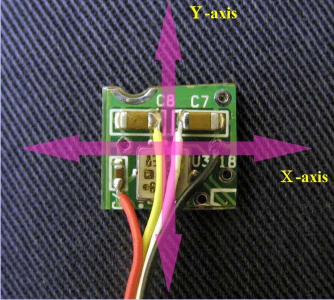
A sensor that records jaw movement
What electromyography records is muscle activity.
It does not record how the jaw moved.
The director therefore introduced an accelerometer, which records movement, into the research on tooth grinding during sleep.
The sensor is a small board about 1 cm square and about 1 mm thick; a thin film inside vibrates with movement, and this vibration is converted into an electrical signal.
It is a device that was used for research.
Recording in clinical practice
In clinical practice, we carry out electromyography for patients for whom we judge it necessary in detailed examinations for TMD.
It is not an examination that we carry out in the same way for everyone.
We also choose the other examinations needed according to symptoms and condition, such as recording tooth contact during sleep (BruxChecker) and autonomic reflex testing (heart rate variability and pupillary light reflex).
Examination duration, number of visits and fees → Fees
What electromyography can and cannot tell us
What it can tell us
- When, and how much, the chewing muscle (masseter muscle) worked (the magnitude of its electrical activity)
- Muscle activity as a percentage of maximum clenching (%MVC)
- Differences in activity between the left and right masseter muscles
- When recorded at the same time as the electrocardiogram, how muscle activity and changes in heartbeat overlap in time
What it cannot tell us, and how we compensate
| What it cannot tell us | How we compensate |
|---|---|
| In which direction, and how far, the jaw moved | In our research, this was recorded with an accelerometer |
| Which teeth make contact during sleep | BruxChecker |
| Jaw joint movement | Cadiax |
| Autonomic nervous activity | Heart rate variability and pupillary light reflex |
| The cause of symptoms such as headaches and stiff shoulders | We do not determine it from muscle recordings alone. Depending on the symptoms, we recommend assessment by a physician |
What electromyography shows is how much the muscles were working at the time of recording.
What the 2026 paper recorded was masseter muscle activity when the occlusal condition was changed temporarily; further research is considered necessary on the relationship with occlusal conditions that have persisted for a long time.
We consider records of muscle activity separately from the cause of each person's symptoms and the effects of their treatment.
Frequently asked questions
- Where are the electrodes attached?
In the study reported in the 2026 paper, they were attached to the skin over the left and right cheek muscles (masseter muscles).
Before they are attached, the skin is wiped with an alcohol swab.
- Can an electromyography recording show the cause of symptoms?
We do not determine the cause of symptoms from electromyography recordings alone.
What electromyography shows is how much the chewing muscles were working at the time of recording.
Depending on the symptoms, such as headaches or stiff shoulders, we also recommend assessment by a physician.
- Why did participants in the research first clench as hard as they could?
To obtain a reference for comparison.
Using the recording of maximum clenching as the reference allows muscle activity at other times to be expressed as a percentage.
- Can electromyography show tooth grinding during sleep?
In the director's research, electromyograms of the masseter muscle were recorded during sleep in a sleep laboratory.
Because jaw movement cannot be recorded by electromyography, the research combined it with an accelerometer.
In clinical practice, where needed, we check tooth contact during sleep with BruxChecker.
Related technologies and treatments
- Heart rate variability (HRV)
- Pupillary light reflex (Iriscorder Dual)
- BruxChecker
- Cadiax
- All the technologies we use
- Temporomandibular disorders & orthodontic treatment that avoids extractions where possible: how treatment proceeds
- Research, publications and conferences
Sources we referred to
For the explanations of electromyography and the accelerometer, we referred to the following published sources (the director's papers). What we describe about our clinical methods is based on the director's posts and explanations.
- Yoshimi H, Suzuki T, Ono Y. Autonomic Responses to Tooth Grinding under Different Dental Occlusal Conditions. Advanced Biomedical Engineering. 2026;15:36-45
- Yoshimi H, Sasaguri K, Tamaki K, Sato S. Identification of the occurrence and pattern of masseter muscle activities during sleep using EMG and accelerometer systems. Head Face Med. 2009;5:7
- Nukazawa S, Yoshimi H, Sato S. Autonomic nervous activities associated with bruxism events during sleep. CRANIO. 2018;36(2):106-112
Consultations and enquiries
Gibakai Medical Corporation
Yoshimi Dental Clinic
Square 2, Room 302 (third floor)
1-29-1 Tsukushino, Machida,
Tokyo 194-0001, Japan
TEL 042-795-1359 FAX 042-795-1398
Clinic days: Mon · Tue · Thu · Fri
10:00–13:00 / 14:30–18:00
A first visit begins with a first consultation (a private, non-insured service; 30 minutes to 1 hour) for examination and diagnosis. Questions about a diagnosis you received elsewhere are also covered within it.
In an emergency, such as being unable to chew or having severe symptoms, please call us on 042-795-1359 to discuss it.